CLOSE X

Blog

Role of accredited biochemical reagents in aneuploidy and pre-eclampsia screening

Role of appropriate NT (nuchal translucency) measurement in aneuploidy screening is well established. Deviation of standard deviation and median NT from that expected, reduces the detection rates and hence continuous auditing of NT measurements is essential for quality assurance (Kagan et al. Ultrasound Obstet Gynecol 2009; 33: 657 – 664).

Similarly, it is important that laboratories analysing biochemical markers for aneuploidy and pre-eclampsia screening, also follow the same stringent criteria which was used during the research to develop the algorithm for screening test. It is necessary that when FMF algorithm (software) is used to calculate the risks, the laboratories should:

  • Use FMF accredited reagents (Delfia, Roche or Kryptor)
  • Ensure internal audits and controls
  • Submit their results for auditing to an external agency like UKNEQUAS for quality assurance.

Deviation from expected MoMs will reduce the detection rates for condition being screened and the efficacy of the test.

Everyone who is offering first trimester screening tests should understand the nuances of the tests being offered and ensure strict adherence to the quality of the tests being done. As an obstetrician, one should ensure NT is measured by FMF certified operator who uses the FMF risk calculation software and biochemical tests are done by laboratories fulfilling the above criteria.

A Case of Split Cord Malformation

Mrs X, 29 years of age

  • Primigravida with 18+6weeks period of gestation
  • First trimester combined screening low risk for aneuploidies
  • Referred to ACFM with diastometomelia in an anomaly scan done elsewhere.

On USG
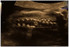
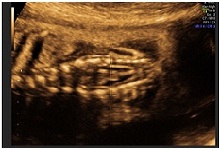
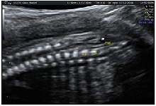
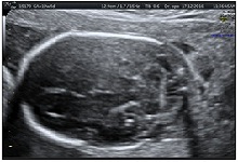
1a1b1c1d2a

Introduction
Diastematomyelia (also termed diplomyelia, pseudodiplomyelia, dimyelia, and split spinal cord malformation syndrome) is a rare developmental deformity in which the spinal cord is separated into two parts by a rigid or fibrous septum. The incidence of diastometomelia is unknown and deformity is often accompanied by abnormal development of the vertebrae.1

Case
One such case was a Primi 18+6 Mrs X who was referred to Apollo Centre for Fetal Fedicine, New Delhi. The anomaly scan at ACFM confirmed the diagnosis of Split cord malformation. Divarication of the fetal spine was seen at the thoracolumbar level(below T 12). Figure 1b. There did not appear to be any mass lesion and the overlying skin was intact. However, the spinal cord appeared tethered, the conus medularis ending at L5 S1 level. Figure 1c. Both the lower limbs were seen to be moving well.

Fetal brain appeared normal. Fetal growth and amniotic fluid was normal. There was no other structural defects nor any markers for chromosomal abnormalities. The couple was explained that cutaneous stigmata(like hairy patch, capillary hemangioma could be seen in 30% of the neonates and is not identifiable on ultrasonography antenatally. They were explained that diastometomelia is a treatable condition but the baby may require postnatal neurosurgery after birth.

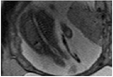
MRI done at Apollo Hospital confirmed the diagnosis of Diastometomelia, figure 2. The couple had a detailed discussion with the neurosurgeon who explained them the prognosis of the baby and the good outcomes of the neurosurgery if at all it will be required. She was send back to her referral obstetrician

The couple decided to continue with the pregnancy and had a follow up with us at 32 weeks. The fetal growth and Dopplers were normal. Good fetal movements were perceived by the mother and were seen on the scan as well.

The woman had an emergency caesarean section at her local hospital at 38 weeks in view of preterm rupture of membranes followed by non reassuring fetal heart rate pattern. The baby weight was 2kg with an APGAR score of 9,9. Post natal MRI showed cord tethering and the baby was planned for surgery at 6 month. However, a repeat MRI at 4 months showed the diagnosis of Diatometomelia type 2(milder form) and the baby was decided to be kept on conservative management. The baby at 6 months of age is doing well.

Discussion
Diastematomyelia is a rare congenital anomaly that results in a longitudinal split of the spinal cord. There are two types of disease entity knwon. Type 1 and Type 2.

In type I, the two hemicords are typically separated by a fibrous, cartilaginous, or osseous septum and reside in two separate dural tubes (Figure 7). Whereas, in Type II split cord malformations both the hemicords lie withina single, non-duplicated, dural tube[2].

The two hemicords typically reunitecaudally, though two coni medullarae may be seenin diplomyelia, an embryologically distinct entity[3].Diastematomyelia is typically associated with vertebralsegmental anomalies.

The majority of patients with diastematomyelia when they grow up are symptomatic, presenting with signs and symptoms of tethered cord, although patients with mild type II may be minimally affected or entirely asymptomatic. Presenting symptoms include: leg weakness, low back pain , scoliosis, incontinence making an individual dependent for life. Another indication of this syndrome is abnormal skin in the lumbosacral region, which may be a hairy patch (hypertrichosis), a pigmented area in the midline, or a dermal sinus[4]

Prenatal Diagnosis: Diastometomelia may go unnoticed by an unexperiened but the presence of an echogenic structure extending from the posterior elements to the posterior aspect of a vertebral body, has high specificity for diastematomyelia[5, 6,].

One third of the patients have an associated spinal dysraphism. The disease has a better prognosis when isolated. So care should be taken to rule out other neural tube defects. Prenatal MRI can be used to confirm the diagnosis of diastometomelia and to rule out other structural defects in the fetus.

Prognosis: The overall prognosis of diastometomelia is good and when indicated, surgical intervention includes decompression of the neural elements with excision of the interposing tissue and reconstruction of the duplicated
dural sacs.

References:
1.Liu W, Zheng D, et al. Characteristics of osseous septum of split cord malformation in patients presenting with scoliosis: a retrospective study of 48 cases. Pediatr Neurosurg 2009; 45: 350–353
2.Pang D, Dias MS, Ahab-Barmada M. Split cord malformation: Part I: A unified theory of embryogenesis for double spinal cord malformations Neurosurgery 1992; 31: 451-480.
3.Cheng B, Li FT, Lin L. Diastematomyelia: a retrospective review of 138 patients. J Bone Joint Surg Br 2012; 94: 365-372.
4.Cheng B, Wang SK, Sun ZC, et al. Analysis of 46 cases of diastematomyelia. Chin J Orthop1996; 16: 97–100.
5.Blondiaux E, Katorza E, Rosenblatt J, Nahama-Allouche C, Lenoir M, le Pointe HD, Garel C. Prenatal US evaluation of the spinal cord using high frequency linear transducers. Pediatr Radiol 2011; 41: 374-383.
6.Glenn OA, Barkovich J. Magnetic resonance imaging of the fetal brain and spine: an increasingly important tool in prenatal diagnosis: part 2. AJNR Am J Neuroradiol 2006; 27: 1807-1814.

Aanchal Sablok MS(ObGyn), Fellow Fetal Medicine (ACFM)

World Prematurity Day

preimage

Magnitude of problem

Around the world, 15 million babies are born prematurely (Less than 37 completed weeks) every year and one million of these will not survive due to complications of preterm birth.
Worldwide 1 in 10 babies are born premature. Sadly, prematurity is the leading cause of death in children under five.

World prematurity day is a global movement to raise awareness about premature birth and the hurdles babies and parents face every day to see their little one survive the day. Many survivors face a lifetime of disability, including learning disabilities and visual and hearing problems.

While World Prematurity Day is an opportunity to call attention to the heavy burden of deathand disability and the pain and suffering that preterm birth causes, it is also a chance to talk about Solutions.

prematurity-blog-apollo-centre-for-fetal-medicine-delhi

The 10 countries with the greatest number of preterm births1:

  • India: 3 519 100 (1 in 7)
  • China: 1 172 300
  • Nigeria: 773 600
  • Pakistan: 748 100
  • Indonesia: 675 700
  • The United States of America: 517 400
  • Bangladesh: 424 100
  • The Philippines: 348 900
  • The Democratic Republic of the Congo: 341 400
  • Brazil: 279 300

In almost all countries with reliable data, preterm birth rates are increasing.Inequalities in survival rates around the world are stark. In low-income settings, half of the babies born at or below 32 weeks (2 months early) die due to a lack of feasible, cost-effective care, such as warmth, breastfeeding support, and basic care for infections and breathing difficulties. In high-income group, almost all of these babies survive.

Addressing preterm birth is now an urgent priority for reaching Millennium Development Goal. Participation from government agencies, NGO’s and private sector is required equally for research, policy making and execution of programs to reduce the toll of preterm birth in high-burden countries like India.

Why does preterm birth happen?

Common causes of preterm birth include multiple pregnancies, infections and chronic conditions such as diabetes and high blood pressure; however, often no cause is identified. There could also be a genetic influence. Better understanding of the causes and mechanisms will advance the development of solutions to prevent preterm birth.

Solution: begins with healthy mother and good antenatal care

  • Counselling on healthy diet, optimal nutrition, personal hygiene and substance abuse
  • Contact with health care professional
  • prematurity-blog-ACFM-delhi

    • Provision of antenatal steroids
    • Essential care during child birth and postnatal period to mother and child
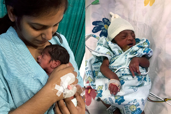
    • Kangaroo mother care- exclusive breast feeding and skin to skin contact
    • Antibiotics to treat neonatal infections

Optimising Use of Antenatal Corticosteroids for Fetal Lung Maturity

Author: Rachna Gupta

It is a well-known fact that antenatal corticosteroids (ACS) reduce perinatal mortalityby reducing the incidence of respiratory distress syndrome (RDS) in premature babies (1).

Cochrane review 2017 reveals that a single course of ACS significantly reduces the incidence of neonatal death by 31%. Meta-analyses have revealed significantly reduced rates of RDS, intraventricular haemorrhage (IVH), necrotizing enterocolitis (NEC), infectious morbidity, need for respiratory support, and neonatal intensive care unit admission with ACS treatment. For the mother, corticosteroid use does not increase the risk of death or chorioamnionitis, although a non-significant risk of puerperal sepsis was found (1). Maximum benefit is after 24 hours to 7 days of completed course.

Two regimes are commonly used:

  • Betamethasone 12 mg I/M 24 hourly 2 doses
  • Dexamethasone 6 mg I/M 12 hourly 4 doses.

Betamethasone is proven to be better than dexamethasone in reducing the neonatal morbidity and mortality, and cause less incidence of chorioamnionitis(1,2). However, as the betamethasone salt available in India is different from that used in international trials (3), same conclusion cannot be drawn, and either dexamethasone or betamethasone may be used depending upon availability.

There is also concern over increased prevalence of undiagnosed maternal infections in India, hence, more trials are needed on safety of use of corticosteroids. One of the trials showed increase in neonatal mortality and maternal infection with use of ACS in low-income and middle-income countries (4).

Use of ACS in late preterm babies (34 – 37 weeks) and early term (37 – 39 weeks) has been a matter of controversy so far and more data are needed to either support or refute the use of ACS in these populations. There are definite short-term benefits (mainly in reducing transient tachypnea of newborn) with some complications like neonatal hypoglycemia and unknown long-term neurodevelopmental outcomes (6).

Studies in animal models and short term studies in human beings have shown that there is increased basal level of cortisol in babies exposed to even single dose of ACS and there is aggravated response to stress (5). Barker’s hypothesis for fetal programming of adult onset of cardiovascular diseases like hypertension, stroke and diabetes also implicates exposure to ACS as a risk factor though this remains to be proven in human studies.
Repeated use of ACS has been linked with autism, neuro-developmental delay, permanent changes in limbic system, midbrain, white matter paucity, cerebral palsy, reduced head circumference, fetal growth restriction, maternal glucose intolerance. All these effects have been more widely studied in animals, and in human beings it is still a speculation, and not proven.
More than 4 repeated doses have been linked with reduced head circumference and fetal growth restriction (7).

Recommendations on basis of current literature:

  • One needs to weigh the potential risks and benefits of ACS. Between 26 – 34 weeks, the potential benefits clearly outweigh the risks and one should not hesitate to give steroids where delivery is anticipated before 34 weeks.
  • The assessment for iatrogenic or spontaneous preterm delivery needs to be more stringent and senior and more than one obstetricians should be involved in decision taking to give steroids at optimal time, so that timing is such that single course is given when delivery is expected before 34 weeks, and the estimated time to deliver is between 48 hours – 7 days.
  • High risk of imminent delivery is strictly defined as (6)

    • Preterm labour with intact membranes and at least 3 cm dilation or 75% cervical effacement
    • Preterm labour with spontaneous rupture of membranes.
    • Expected preterm delivery for any other indication either through induction or caesarean section between 24 hours and 7 days, as determined by the obstetrical provider.
  • Single course of ACS may be repeated if gestational age is less than 34 weeks, more than one week has elapsed and delivery is strictly expected between 24 hours to 7 days.
  • Between 34 – 37 weeks, there are short-term benefits with unknown long-term side effects, and lung maturity tests (like QuantusFLM) may be useful to assess the need for ACS.
  • There is not enough data to support use of ACS after 37 weeks either for planned C-section or normal delivery (8,9).
  • Elective C-section should be postponed to 39 weeks as much as possible as risk for respiratory distress keeps reducing and is minimum after 39 weeks.

References:

  • Roberts D, Brown J, Medley N, Dalziel SR, Antenatal corticosteroids for accelerating fetal lung maturation for women at risk of preterm birth, Cochrane Database Syst Rev. 2017 Mar 21;3:CD004454.
  • Baud O, Sola A; Corticosteroids in perinatal medicine: How to improve outcomes without affecting the developing brain? Semin Fetal Neonatal Med. 2007 Aug;12(4):273-9. Epub 2007 Mar 21. Review.
  • Use of Antenatal Corticosteroids in Preterm Labour; Operational Guidelines June 2014; Ministry of Health and Family Welfare, Government of India.
  • Fernando A et al; A population-based, multifaceted strategy to implement antenatal corticosteroid treatment versus standard care for the reduction of neonatal mortality due to preterm birth in low- income and middle-income countries: the ACT cluster- randomised trial; Lancet. 2015 February 14; 385(9968): 629–639.
  • Waffarn F, Davis EP; Effects of antenatal corticosteroids on the hypothalamic-pituitary-adrenocortical axis of the fetus and newborn: experimental findings and clinical considerations; Am J Obstet Gynecol. 2012 Dec;207(6):446-54-
  • Beena D. Kamath-Rayne et al; Antenatal corticosteroids beyond 34 weeks gestation: What do we do now? Am J Obstet Gynecol. 2016 Oct;215(4):423-30.
  • Michael W. Church et al;Repeated courses of antenatal corticosteroids: Are there effects on the infant’s auditory brainstem responses?NeurotoxicolTeratol. 2010; 32(6): 605–610.
  • ACOG Committee Opinion number 713, August 2017
  • NICE guideline (NG 25) Preterm labour and birth (published Nov 2015)

Congratulations Dr Vimla Dahiya from Sonepat!

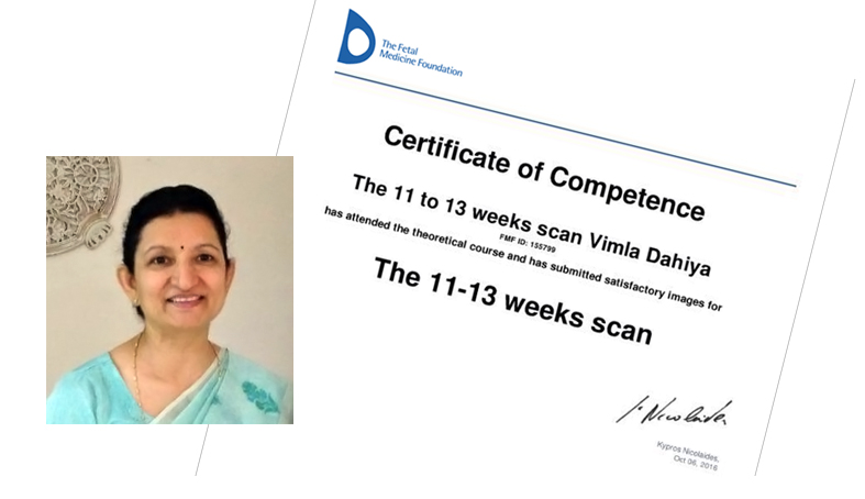
We are proud of Dr Vimla Dahiya, obstetrician from Sonepat, who after attending FMFI outreach programme held in Sonepat on 7th Feb 2016, was determined to get her FMF-UK certification, and with persistence and guidance from Dr Rachna Gupta (sec, outreach programme) obtained the certification on 7th Oct 2016. We expect more and more people to achieve this, to bring uniformity and quality to nuchal scans, which is the aim of our outreach programmes.

vimla-dahiya-certi

Rubella infections

As part of the FMFi- Look for life education outreach, we are starting a monthly newsletter covering subjects which will help towards addressing the problems of perinatal mortality and still birth. The first of these is fetal infections of which the most preventable cause of severe neonatal morbidity is congental rubella infections.

Universal rubella immunization is part of the Govt of India’s childred immunisation programme but nevertheless any woman who is planning pregnancy or is seen at any time during pregnancy has to have her IGG Rubella status checked. ( please note that it is important to not just do an IGM whenever infection screening is done and both IGG and IGM values need to be checked so that correct advice can be given.

Rubella, also called German measles, is a disease of childhood . In the absence of pregnancy, it is usually clinically manifested as a mild self-limited infection.During pregnancy, however, the virus can have potentially devastating effects on the developing fetus. It has been directly responsible for inestimable wastage and for severe congenital malformations.

Recommendations

  • The risk of congenital defects has been reported to be 90% when maternal infection occurs before 11 weeks of gestation, 33% at 11–12weeks, 11% at 13–14 weeks, 24% at 15–16 weeks, and 0% after 16 weeks.7.Since the effects of congenital rubella syndrome vary with the gestational age at the time of infection, accurate gestational dating should be established, as it is critical to counselling. (II-3A)
  • The diagnosis of primary maternal infection should be made by serological testing. (II-2A) The presence of a rubella
    • A fourfold rise in rubella IgG antibody titre between acute and convalescent serum specimens
    • A positive serologic test for rubella-specific IgM
    • A positive rubella culture (isolation of rubella virus in aclinical specimen from the patient)
  • Serologic studies are best performed within 7 to 10 days after the onset of the rash and should be repeated two to three weeks later.
  • Screening to determine the antibody status of all pregnant women to determine susceptibility. Providing programs to ensure postpartum immunization of non-immune women before they are discharged from the hospital.
  • Women who have been inadvertently vaccinated in early pregnancy or who become pregnant immediately following vaccination can be reassured that there have been no cases of congenital rubella syndrome documented in these situations. (III-B)
  • Women wishing to conceive should be counselled and encouraged to have their antibody status determined and undergo rubella vaccination if needed. (I-A)
  • Congenital defects and late manifestations of rubella infection:
    Present at birth Late manifestations : Audiologic anomalies (60–75%) ,Sensorineural deafnessCardiac defects (10–20%) – Pulmonary stenosis , Patent ductus arteriosus, Ventricular septal defectOphthalmic defects (10–25%) –Retinopathy , Cataracts , Microphthalmia , Pigmentary and congenital glaucoma

    Central nervous system (10–25%) – Mental retardation , Microcephaly ,Meningoencephalitis

    Others – Thrombocytopenia , Hepatosplenomegaly ,Radiolucent bone disease ,Characteristic purpura (Blueberry muffin appearance) , Diabetes mellitus , Thyroiditis ,Growth hormone deficit , Behavioural disorder

    (SOGC Guidelines 2008)

FMFIndia academic activities

FMFIndia is dedicated to teaching obstetricians and sonologists about the current standard of antenatal screening protocols and .

  1. 2 week ultrasound programme

The following doctors from all across India have undergone the 2 week ultrasound programme at ACFM under the aegis of FMFI.

2013

  • Dr Divya Aggarwal Lucknow, UP
  • Dr Vandan Jain Rohtak, Haryana
  • Dr Savita Dagar New Delhi
  • Dr Broti Basu Calcutta, West Bengal
  • Dr Kanchi Khurana Chandigarh, Haryana
  • Dr Shivali Mittal Palwal, Haryana
  • Dr Sophia Keshi Imphal, Manipur

2014

  • Dr Jaya Chawla New Delhi
  • Dr Madhushree Ray Naskar Calcutta, West Bengal
  • Dr Usha Gupta Bhatinda, Punjab
  • Dr Pradeep Ingale Ahmadnagar, Maharashtra
  • Dr Neelu Yadav Patna, Bihar

2015

  • Dr Uzma Firdaus Srinagar, Kashmir
  • Dr Disha Bansal New Delhi
  1. 6 month Ultrasound Training Programme
  • Dr Anjali Solanki at Rainbow Hospital, Hyderabad
  • Dr Setu Chhabra at ACFM, New Delhi
  1. CMEs and Workshops
  • 11th Nov, 2012 CME, World Prematurity Day
  • 20th July, 2013 Perceptor Workshop on Fetal Echocardiography, Prof Lindsay Allen
  • 9th Sept, 2013 Maternal Fetal Medicine Symposium Serum
  • 27th Oct, 2013 GE Obstetric Doppler workshop
  • 9th June, 2014 Obstetric Doppler Workshop
  • 7th June, 2014 Maternal Serum Screening Study Day
  • 22nd Jan, 2015 Fetal Cardiac Screening Workshop
  1. Awareness Activities
  • 2nd Dec, 2013 International Day of People with Disability
  • 7th Dec, 2013 World TTTS Day
  • 21st Mar, 2014 World Down Syndrome Day
  • 2nd Dec, 2014 International Day of People with Disability
  • 17th Nov, 2014 World Prematurity Day

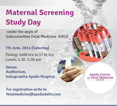
Maternal Screening Study Day, CME organised by FMFIndia

We are pleased to announce ‘Maternal Screening Study Day’ CME under the aegis of subcommittee Fetal Medicine, AOGD

maternal screening

The Highlights:
– First Trimester Screening – standard of care
– Too Late for First Trimester – What to do next?
– Emerging trends in Screening for Aneuploidy
– Clinical Case Scenarios

Also included are a Post CME quiz and Q & A session.

Venue: Auditorium, Indraprastha Apollo Hospital
Date: 7th June, 2014 (Saturday)
Timing: 1400 hrs to 1730 hrs

View programme details at www.fetalmedicineindia.in/screeningcme.pdf

Send in your registration requests, questions to fetalmedicine@apollodelhi.com or +91 9899092076

India losing fewer infants but still short of target

A modest yet consistent decline in the infant mortality rate, especially in six problematic states, is one of the key features of the latest data from the Sample Registration System. Nationwide, the IMR has dropped by three points from 47 infant deaths per 1,000 live births to 44, according to the October 2012 SRS bulletin. It has dropped to 48 from 51 in rural areas , and to 29 from 31 in urban areas, as compared to the previous count.

The comparison is between 2010 and 2011 for larger states, and between two three-year samples for smaller states and Union Territories.

Despite the drop, the prevailing mortality rate remains much higher than the target set under the 11th five-year plan  30 per 1,000 live births by 2012. Under the 12th five-year plan, India hopes to reduce the mortality rate to 25 by 2017.

Health ministry officials concede it is early days yet and a lot needs to be done, but find the consistent drop encouraging. Dr Ajay Khera, deputy commissioner (child health and immunisation) in the ministry, told The Indian Express that a three-point drop in a majority of states is a healthy sign.

In six states  Bihar, Orissa, Uttar Pradesh, Haryana, Punjab and Sikkim  the decline by four points is an achievement,” Khera said. States such as UP and Bihar continue to face major problems such as kala azar and pneumonia. Madhya Pradesh remains disappointing with an IMR of 59/1000, highest of all states, followed by UP and Orissa at 57/1000. All these states have, however, improved since last count.

The data shows 13 states as having reduced their IMRs by three points. These include Maharashtra, Madhya Pradesh, Gujarat, Andhra Pradesh, Assam, Jharkhand, Karnataka and some of the northeastern states.

There has been a drop of two to three points every year. Goa and Manipur have the lowest IMR, 11, followed by Kerala with 12.

The SRS figures are the only national data providing annual estimates, since 1964, on fertility and mortality indicators. The assessment is spread across all states and UTs, covering 1.5 million households and 7.35 million people.

Khera said it is crucial to ensure diarrhoea and pneumonia management for newborns. There are several challenges and it is crucial that the government’s schemes and welfare measures reach all children, said Deepti Agarwal, child health consultant with the central government.

As per WHO estimates, the main causes of child mortality in the age group 0-5 in India are neonatal mortality , pneumonia, diarrhoea, measles and injuries. Neonatal mortality contributes to about two-thirds of infant deaths and nearly half of under-5 deaths. The focus has been on newborn care and, according to Khera, one of the key strategies is the integrated management of neonatal and childhood illness programmes. It is being implemented in more than 400 districts and nearly five lakh health personnel have been trained.

Home-based newborn care is a recent initiative, where at least eight lakh Accredited Social Health Activists under the NRHM programme will be trained. ASHAs will visit all targeted newborns according to a specified schedule.

Special Newborn Care Units at each district hospital with specialised equipment have been earmarked. There are 395 SNCUs today, set up in first referral units and community health centres. These units provide services that include resuscitation, warmth, early initiation of breastfeeding and prevention of infection. And newborn Care Corners are special corners within the labour room where support for optimal management of a newborn is provided.

In Maharashtra, Dr V K Rokade, assistant director in the family welfare bureau, told The Indian Express that the target of reducing infant deaths to 17 per 1,000 live births had not been achieved in 2011, but a three-point drop (28 to 25) is a first for the state that now aims to bring it down to 21 by 2017. A total 1,029 newborn care corners at primary health centres, units at 23 district hospitals, 146 newborn stabilisation units at rural hospitals with paediatricians and trained staff, and a nutrition mission programme have contributed to the state’s campaign to bring down infant deaths.

-Reproduced from The Indian Express online Website