Take home message – Webinar on Fetal Alcohol Spectrum Disorders (FASD)

Featured

Discussants and speakers –

  • Prof Gregor Kasprian
  • Dr Sumathi Natarajan
  • Dr Patric Kienast
  • Dr Marlene Stuempflen
  • Dr Hemlata Bhatia
  • Dr Mainsha Kumar
  • Dr Saurabh Chopra
  • Sophie Harrington 

Hosts – Dr Anita Kaul, Dr Rachna Gupta, Dr Akshatha Prabhu

Study presented by Dr Marlene Stuempflen – 

Stuempflen M, et al. Fetal MRI based brain atlas analysis detects initial in utero effects of prenatal alcohol exposure. Cereb Cortex. 2023 May 24;33(11):6852-6861. doi: 10.1093/cercor/bhad005 

Presentation by Sophie Harrington – Importance of FASD, work being done by NOFASD Australia and Sharing Lived experience of FASD

Take home messages – 

Periconceptional counseling

a) No amount of alcohol is considered safe to be consumed during pregnancy or breastfeeding. 

b) It is necessary to ask history of alcohol intake to every woman and provide information and leaflets for awareness of FASD, however uncomfortable it may be for the clinician and the woman.

Counseling if exposed to alcohol:

a) In the women exposed to alcohol – there is 1/13 chance of child being affected with FASD, and 1/67 chance of being severely affected.

b) Early diagnosis of FASD is beneficial for better management and outcome of affected children, appropriate diagnosis and support services are needed to guide and support families.

Spreading Awareness:

Alcohol is general part of the society and one doesn’t need to be ashamed or feel guilty of accepting the fact that they take alcohol, no judgement or bias should affect the counselling and care.It is not just the woman but also the partner who needs to understand the significance of FASD, to be able to support alcohol abstinence in women planning pregnancy. 

Antenatal Diagnosis of FASD:

There are no well proven ultrasound findings of FASD, however, very severe affection can lead to corpus callosal dysgenesis which may be picked up on ultrasound. Fetal MRI as a routine cannot pick up subtle cases of FASD, the current study used research tools to look at the differences in periventricular zone and corpus callosal volumes, these tools may be available in future once automated based on large studies.

Further research is needed in certain areas – 

a) Role of paternal alcohol intake in the periconceptional period

b) How much time of abstinence from alcohol is needed before planning pregnancy

c) Better designed studies to correlate alcohol intake and effects in children, according to timing and amount of alcohol intake.

d) Tools for early diagnosis and support services for affected families. 


Recording of the Webinar

Fetal Echocardiography-Simplified

‘Fetal echocardiography-simplified’, a book that takes you through the technique of fetal echo, applied embryology and salient features of various cardiac defects, in an easy language, illustrated by line diagrams and corresponding USG images in 2D, Doppler and 3D, to serve a ready reference in your busy clinic, is authored by Dr Nupur Shah, a past ACFMite, who is currently working as consultant fetal medicine and genetics in Baroda.

The book also features special chapters on ARSA and left brachiocephalic vein.

It was launched by Dr Anita Kaul, Director and head, ACFM on 24th March 2023.

It is available worldwide on :

India-  https://www.amazon.in/dp/1636408486?ref=myi_title_dp

https://www.flipkart.com/fetal-echocardiography-simplified/p/itmf6d10382e6f17?pid=9781636408484

USA – https://www.amazon.com/dp/B0BZ8S1H9L

UK – https://www.amazon.co.uk/dp/B0BZ8S1H9L

Germany – https://www.amazon.de/dp/B0BZ8S1H9L

Brazil – https://www.amazon.com.br/dp/B0BZ8S1H9L

Canada – https://www.amazon.ca/dp/B0BZ8S1H9L

Australia – https://www.amazon.com.au/dp/B0BZ8S1H9L

France – https://www.amazon.com.fr/dp/B0BZ8S1H9L

Mexico – https://www.amazon.com.mx/dp/B0BZ8S1H9L

Spain – https://www.amazon.es/dp/B0BZ8S1H9L

Italy – https://www.amazon.it/dp/B0BZ8S1H9L

Netherland – https://www.amazon.nl/dp/B0BZ8S1H9L

WZ research webinar summary June 2022

Prenatal Invasive Testing at a Tertiary Referral Center in India: A Report of 433 Cases Under a Single Operator.

The Journal of Obstetrics and Gynecology of India – Jan 2022
Speaker – Dr. Vandana Bansal
Moderator – Dr. Anita Kaul

The study looked at indications for invasive testing, positive predictive value according to the indication,incidence of second prick, culture failure, or miscarriage within 4 weeks of the procedure.

  1. Maximum number of procedures were due to being screen positive for chromosomal problems. According to the study the overall positive predictive value (PPV) of a screen positive test for chromosomal aneuploidies was 6.7%. The PPV was very low (2.7%) for isolated biochemistry screen positive. Dr. Anita Kaul highlighted that this could be due to patients being referred for invasive testing based on serum biochemistry reports from different labs many of whom might not be adhering to strict FMF protocols for testing and auditing. Therefore, the need to do biochemistry from standardized labs and use only combined risk results rather than isolated biochemistry results for counselling was emphasized.
  2. The study had no procedure related losses. However; it was astutely pointed by Dr. Anita Kaul that this could reflect the author’s personal expertise given her extensive experience and might not apply to all. Also Dr. Anita Kaul recommended that pre term deliveries post procedure could have been included in the outcomes analyzed.

Preserved Blood Spots Aid Antenatal Diagnosis of Citrullinemia Type-1
Journal of fetal medicine – June 2021

Speaker – Dr. Shruti Bajaj
Moderator – Dr. Divya Agarwal

The case report highlighted the importance of DNA storage in case of history of neonatal death with suspected inborn error of metabolism.

  1. Isolated biochemistry-based diagnosis of IEM is not enough to aid couples in reproductive genetic counseling. This has to be coupled with a genomic diagnosis.
  2. Dried blood spots can be a source of DNA when appropriately stored for many years.
  3. DNA banking will go a long way in aiding diagnosis even if index case is no longer available. Therefore obstetricians and neonatologists need to be made aware about DNA banking methods.

Especially in fetuses that are being terminated for malformations and neonatal deaths where cause has not yet been firmly established.

Disclaimer:

The papers presented in the research webinars strictly reflect the author’s opinions and practices. FMFI is a neutral platform for scientific discussions and does not endorse or refute these opinions.

Autistic Pride Day – 18 June 2022

As per recent data ASD is reported in 1:59 children. Autism spectrum disorder (ASD) is a neurodevelopmental disorder characterized by deficits in social communication and social interaction and the presence of restricted, repetitive patterns of behavior, interests, or activities present during early periods of development that negatively impact social, occupational, or other domains.

Autism affects 2 key areas:

  • Defective Social interactions and social communication skills
  • Restricted, repetitive patterns of behavior, interests, or activities

These symptoms cause clinically significant impairment in social, occupational, or other important areas of current functioning.

These disturbances are not better explained by intellectual disability (intellectual developmental disorder) or global developmental delay. However, autism and intellectual disability can coexist together

The average age of diagnosis is 5 years; however, early warning signs can be observed in infancy.

Early intervention based on the principles of ABA can improve the functioning of chil- dren with ASD by addressing behavioral deficits and excesses.

  • Interventions based on principles of applied behavior analysis (ABA) can effectively reduce social skills deficits following training.
  • Approaches to language development and communication based on ABA incorporate a functional approach to language and can effectively increase verbal and nonverbal communication skills.

The cause of ASD is unknown, but research supports genetic and environmental factors.

Common genetic causes for autism include Fragile X syndrome, Tuberous sclerosis and 22q11 deletion. Hence, Chromosomal microarray and Fragile X screening are the thefirst tier test in cases of autism.

In the absence of specific genetic test results, only recurrence risk estimates can be made; the recurrence rate with one previously affected sibling is around 10–15%. If there are two affected siblings in the family, the estimated rates predicted by a theoretical model are around 50% and 12% in subsequent newborn boys and girls, respectively . The recurrence rate varies as a function of the gender of the previously affected sibling, with higher recurrence rates in the case of a female affected sibling

The increasing prevalence of ASD and substantial economic and collateral costs at the individual, familial, and societal levels necessitate attention to interventions.

Dr. Seema Thakur
MS (Obs &Gynae), DM (Med Genetics)
Senior Consultant Genetics and Fetal Diagnosis

World Sickle Cell Day – 19 June 2022

Sickle cell disease(SCD) is one of the most common monogenic disorders globally with an autosomal recessive inheritance1. James Herrick, a physician first described the characteristic sickle shaped red cells in a medical student from Grenada in 1910.The disease results from a single base A>T mutation in the triplet encoding the sixth residue of the β-globin chain, leading to a substitution of valine for glutamic acid and the abnormal haemoglobin S (HbS).

The primary pathophysiology is based on the polymerization of deoxyHbS with formation of long fibers within the RBCs causing a distorted sickle shape which eventually leads to increased haemolysis and vaso-occlusion of sickle red cells.

The sickle cell gene is predominantly seen among the tribal populations in India, many of whom live in remote hilly regions . Carrier frequencies range from 1 to 35% in many commun- ities. The disease in India occurs predominantly in a central belt stretching from Eastern Gujarat, Maharashtra, Madhya Pradesh, Chhattisgarh, to Western Odisha with a smaller focus in the Nilgiri Hills of northern Tamil Nadu and Kerala in southern India.

A recent calculation using currently available data indicates that 50% of the total AS and SS neonates are born in only three countries, Nigeria, India and DR Congo. For SS neonates, the estimate for India was 42 016, which corresponds to 88% of the homozygous cases in Asia [14&]. This emphasizes the need for a national registry for an accurate estimation.

Prenatal diagnosis is indicated if both husband and wife are sickle cell trait. Risk of sickle cell is 25% and prevention is possible by CVS at 11-12 weeks of pregnancy
Sickle cell anemia management can be considered in two categories, health maintenance, and management of complications.

The goal of health maintenance is to screen and identify risk factors and early signs of complications. There is evidence that pneumococcal vaccination, penicillin prophylaxis (early infancy until at least age five and education of the management of fever have considerably reduced mortality and morbidity from invasive infections.

Routine screening with transcranial Doppler (TCD) of large intracranial blood vessels may predict the risk of stroke in children with sickle cell

Management of sickle cell complications is tailored to the type of complication. The management consists of rapid pain assessment, early initiation of analgesic therapy, and maintaining this analgesia (consider PCA pump) and hydration until there is symptom relief. For most patients with mild pain, acetaminophen or NSAIDs may suffice, but for moderate and severe pain, opiates with or without NSAIDs are indicated
Among patients who have frequent and severe complications, hydroxyurea has been shown to offer significant benefits. Hematopoietic stem cell transplantation (bone marrow transplantation) shows great promise as a cure for sickle cell disease. Antihistamines (diphenhydramine, hydroxyzine) may reduce itching caused by the opiates and may have some opioid-sparing effect. For acute chest syndrome, empirical antibiotics (cephalosporin and macrolide), adequate analgesics, oxygen supplementation, simple or exchange transfusion, beta-agonist inhalations, and incentive spirometry may be needed.

Dr. Seema Thakur
MS (Obs &Gynae), DM (Med Genetics)
Senior Consultant Genetics and Fetal Diagnosis

TRIVIA

Little People of America (LPA) is a nonprofit organization that provides support and information to people of short stature and their families.LPA is dedicated to improving the quality of life for people with dwarfism throughout their lives while celebrating with great pride Little People’s contribution to social diversity.  LPA strives to bring solutions and global awareness to the prominent issues affecting individuals of short stature and their families.


The Osteogenesis Imperfecta Federation Europe (OIFE) is an umbrella association for organizations dealing with the rare condition Osteogenesis Imperfecta (OI) also known as brittle bone disease. The federation was established in 1993 by six founding OI-organizations and is registered as a non profit in the Netherlands.


Surgical procedure called “rodding” is frequently considered for people with OI. This treatment involves inserting metal rods through the length of the long bones to strengthen them and prevent and/or correct deformities.


Famous characters in Hindu mythology that were short in stature and did great things -Agastya,Vamana,Kubera,Siva Ganas

trivia-banner

There is evidence that OI has affected people since ancient times. It has been recognized in an Egyptian mummy of an infant from about 1000 BC. The mummy is currently in the British Museum in London, England The partially reconstructed skull is the remains of an Egyptian mummy, origins dated circa 1000 BC. Thought to be remains of a monkey, a first further investigation by paleopathologists revealed that these findings are more consistent with the remains of an infant affected by osteogenesis imperfecta


possible to promote muscle and bone strength, which can help prevent fractures. Swimming and water therapy are common exercise choices for people with OI, as water allows independent movement with little risk of fracture


Nicolas de Malebranche (born 1638) is thought to be the first person to attempt to describe the clinical features of OI


Greek teller of fables Aesop described as one whom nature had gratified with an ingenious mind, he was endowed with a large head, bowed legs and a large belly.


Diastrophic dysplasia affects bone and cartilage development. (“Diastrophism” is a general word referring to a twisting.)


Campomelic dysplasia, the name is derived from the Greek work campo (or campto), meaning bent, and melia, meaning limb


Thanatophoric means “death bearing” in Greek as the children born with were either stillborn or died immediately after birth .


Osteogenesis imperfecta literally means “bone that is imperfectly made from the beginning of life.”


A Viking leader who lived in the 9th century, Ivar Ragnarsson “Ivar the Boneless,” probably had OI. He is reported to have been a very wise leader and a very fierce warrior who had to be carried into battle on a shield because his legs were so soft.

A case of timely prenatal testing of current pregnancy based on evaluating an index case with uncertain diagnosis.

Authors:Dr Sumitra Bachani,* Dr Suchandana Dasgupta ** * Professor & Senior Specialist,** NBE Fellow Maternal-Fetal Medicine Vardhman Mahavir Medical College & Safdarjung Hospital

Introduction: There are more than 400 known types of generalized skeletal dysplasia. The estimated prevalence is 2-7 in 10,000 live births.1 The genetic etiology of an increasing number of cases with skeletal dysplasia is now known, making an early diagnosis possible. In the case of a previously affected pregnancy witha definitive geneticdiagnosis,prenatal genetic testing can thenbe offered. Inheritance patterns can vary; therefore, Genetic counselling and appropriate Next-generationsequencing (NGS), such as Clinical exome or Whole exome sequencing (WES) modalities,help investigate affected pregnancies and arrive at a diagnosis.

We report such a case where a correct diagnosis of the proband facilitated a timely prenatal test in the subsequent pregnancy.

Clinical findings and relevant history: A 34 year G3P2L2 with 13 weeks of gestation presented with one affected female child of 7 years with a history of delayed dentition, inability to walk, multiple fractures, and bowing of both legs since she was 8 months old. (Fig 1A-1D) On investigation of the girl child’s serum calcium, phosphate, and parathyroid hormone (PTH) levels were normal, but alkaline phosphate (ALP) was high (336 IU/L). A bone biopsy was performed at 10 months of age, and the histopathological features were consistent with fibrous dysplasia. Magnetic Resonance Imaging (MRI) at one year of age revealed a fracture of the lower diaphysis of the femur with mild angulation. She underwent multiple surgeries for repeated fractures and later planned for deformity correction for bowing of her legs. However, no definitive diagnosis was made, and probable conditions like fibrous dysplasia, osteogenesis imperfecta and metabolic rickets were considered. She has another healthy male sibling of 10 years of age. It was consanguineous marriage, and all were spontaneous conceptions. Three generation pedigree was not significant

IMAGES
Fig 1A: Current photograph of index child; Fig 1B: X-ray (A.P. view) of bilateral pelvis with bilateral femur bowing and bilateral femur meta-diaphysis fracture (age 10 months); Fig 1C: X-ray (lateral view) showing anterolateral bowing of bilateral femur (age 10 months); Fig 1D: X-ray (A.P. view) showing left humerus bowing with fracture and crowding of ribs at 4 years of age

Diagnostic assessment: The current pregnancy was confirmed by ultrasound at 10 weeks gestation. A nuchal translucency scan was done at 12 weeks, and Non-Invasive Prenatal Screening (NIPS) was also done, which was low risk for aneuploidy. With genetic counselling and pretest counselling, the proband was offered Whole-exome sequencing (WES). The WES revealed that the proband was homozygous for pathogenic variant consistent with osteogenesis imperfecta type VI having autosomal recessive inheritance. (Fig2)

table
Fig 2: Whole Exome Sequencing of index child showing mutation of SERPINF1 gene.
At 17 weeks, amniocentesis was performed with Sanger sequencing, which revealed the fetus to be heterozygous for O.I. VI. (Fig 3). The couple were counselled post-test that the baby would not be affected but a disease carrier.

IMAGE

The couple was offered carrier screening, and both partners were detected to be heterozygous carriers for the same condition. (Fig 4)

IMAGE

Follow up and outcome: The Anomalyscan done at 18 weeks was normal, and follow up growth scans done at 32 and 36 weeks were also unremarkable. The mother had an uneventful labour and delivered a 3.1 kg male baby at term. The postnatal period was uneventful. The baby is of 9 months of age and doing well to date.

Discussion: Osteogenesis imperfecta (O.I.), also known as brittle bone disease, is a genetic disorder of connective tissues caused by an abnormality in the synthesis or processing of type I collagen. A total of 17 types of subgroups have been identified depending upon the genetic mutations.2

In the presence of consanguinity, autosomal recessive conditions shouldbe considered. Type VI mutation involvesthe SERPINF1 gene; characteristic histological presentation includes lamellar bone with fish scale pattern under a polarized light microscope and severe mineralization defects. It presents with moderate to severe skeletal manifestations, normal sclera, and absence of dental involvement.3 Byers et al. formulated a guideline for genetic evaluation in suspected cases of O.I.4 They suggested that if in the first year of life there are frequent unexplained fractures that are non-accidental, it may raise suspicion of O.I. On clinical examination, if an infant with unexplained fractures has few features of O.I., as may be the case with O.I. type I, IV, V and VI, it may be difficult to confirm or exclude the diagnosis based on the history of fractures, family history, and physical examination alone, particularly in the 0–8 months age group. Herein lies the role of genetic history and phenotype identificationin ascertaining the pathology.

Moldenhauer et al. reported two Brazilian families from a small city called Bueno Brandao in Southeast Brazil with a severe deforming form of autosomal recessive O.I., in which WES identified a novel 19-bp homozygous deletion in exon 8 of SERPINF1. All affected individuals in this study had their first fracture approximately at 1 year (except one who had their first fracture at 5 months). None of them had dentinogenesis imperfecta, and only one had mild blue sclera. All affected individuals had a homozygous 19-bp deletion in exon 8 of SERPINF1 (c.1152_1170del; p.384_390del)

In their study, Francis H et al. described 8 patients initially diagnosed with O.I. type IV who shared uniquecharacteristics. Fractures were first documented between 4 and 18 months of age.Patients with O.I. type VI sustained more frequent fractures than patients with O.I. type IV. Sclerae were white or faintly blue, and dentinogenesis imperfecta was uniformly absentAll patients had vertebral compression fractures and no radiological signs of rickets. Lumbar spine bone mineral density was low and similar to age-matched patients with O.I. type IV. Serum alkaline phosphatase levels were elevated compared with age-matched patients with type IV OI (4096145 U/litre versus 295695 U/litre; p<0.03 by t-test). Mutation screening of the coding regions and exon/intron boundaries of both collagen type I genes did not reveal any mutations and type I collagen protein analyses were normal. They proposed to name this disorder “O.I. type VI.” &nsp;

Thus in the current case, the proband’s clinical presentation was not classical of O.I., and she had an unaffected sibling. Although there was a suspicion of O.I., no genetic counselling or confirmatory genetic tests were done. NIPS had been done, which was irrelevant and not cost-effective for this condition. It is essential to thoroughly investigate the affected case with appropriate tests. Otherwise, the window period for investigations and diagnosing the disease becomes narrow if the woman conceives again, as was in the current case. The couple was thus helped with the decision regarding the current pregnancy. Also, they were both detected to be the carriers and understood the need for prenatal tests in every pregnancy. The strength of the case workup was the timely assessment of the proband and the role of Genetic counselling and appropriate testing. The limitation is that we had to rely on WES to diagnose the condition as the presentation was not classical.

Reference:

  1. Orioli IM, Castilla EE, Barbosa-Neto JG. The birth prevalence rates for the skeletal dysplasias. J Med Genet. 1986;23(4):328-32.
  2. Van Dijk FS, Byers PH, Dalgleish R, Malfait F, Maugeri A, Rohrbach M, et al. Best practice guidelines for the laboratory diagnosis of osteogenesis imperfecta. Eur J Hum Genet. 2012;20:11–19.
  3. Subramanian S, Viswanathan VK. Osteogenesis Imperfecta. StatPearls; 2022.
  4. Byers P, Krakow D, Nunes M. et al. Genetic evaluation of suspected osteogenesis imperfecta (O.I.). Genet Med 2006;8:383–88.
  5. Moldenhauer Minillo R, Sobreira N, de Fatima de Faria Soares M, Jurgens J, Ling H, Hetrick K, et al. B: Novel Deletion of SERPINF1 Causes Autosomal Recessive Osteogenesis Imperfecta Type VI in Two Brazilian Families. Mol Syndromol 2014;5:268-275.
  6. Glorieux, F.H., Ward, L.M., Rauch, F., Lalic, L., Roughley, P.J. and Travers, R. (2002), Osteogenesis Imperfecta Type VI: A Form of Brittle Bone Disease with a Mineralization Defect. J Bone Miner Res, 17: 30-38.

BINDER PHENOTYPE:Decision Making at a Tough Crossroad

Vikram N S, Savita shirodkar, SrimathyRaman, Lathavenkataram

Fellow in Foeto-Maternal medicine, 2 Consultant, 3Director, South Bangalore Obstetrics and Gynaecology Doctors and associates (SBOGYN team), Rangadore Memorial Hospital, Bangalore- A Unit of Sri Sringeri Sharada Peetam Charitable Trust, (Affiliated to RGUHS, Karnataka)

Introduction
Binder phenotype with stippling results in hypoplasia of the nasal pyramid (also called maxillonasal dysplasia).If other anomalies are visible, the risk that Binder syndrome is part of another syndrome is high.
Curry et al.in 1993.first published the association between Chondrodysplasia punctate (CDP)with characteristic Binder’s phenotype and maternal autoimmune connective tissue disorder.

Mixed connective tissue disease (MCTD) is characterized by high titre antibodies to U1-ribonucleoprotein (RNP).Even thoughCDP fetuses,which are associated with maternal autoimmune disorders, have a comparatively better prognosis, the inability todifferentiatethis underlying causeon the part of the clinician may result in an unnecessary termination of the pregnancy.

CASE REPORT: Relevant history and Clinical findings 28-year-old primigravida booked at six weeks of pregnancy.She was a known case of mixed connective tissue disorder with Anti Ro, Anti Sm and U1RNP antibody positive and was on Hydroxychloroquine, Azathioprine and steroids fortwo years.She was in remission and was seen preconceptionally andcounselled about the condition.She was being jointly managed with the rheumatologist.Her first-trimester screening was normal.

Diagnostic assessment: Second-trimester anomaly scan done at 19 weeks showed Binder’s Phenotype with stippling of the epiphysis of long bones and coccyx with suspicion of Chondrodysplasia punctata in the fetus.

Intervention: The patient was counselled that she needs further evaluation with anamniocentesis and microarray/exome.However, she opted to continue the pregnancy without invasive /genetic testing.In the backgroundof MCTD, the possibility that this fetus had CDP was high.

Follow up: Her maternal condition remained stable
She was followed up with mechanical PR interval monitoring for the diagnosis of AV block because of her Ro, La positivity.Follow up scan done every fortnight continued to show the bony stippling (femur epiphyseal and coccygeal stippling )but did not show any progress of the lesion.

BINDER

She delivered a baby of 3kg by c section.Post-delivery scans did not show any additional anomaly.

Outcome:The infant attained normal growth and development as per WHO growth charts and development guidelines.

DISCUSSION
Review of literature

The concept of MCTD as a separate immune-mediated connective tissue disease was first introduced by Sharp et al.2>40 years ago, but there is still no consensus regarding the disease definitions, classification, criteria or the relationship with autoimmune conditions.MCTD may begin with any clinical manifestation associated with SLE, systemic sclerosis, polymyositis or rheumatoid arthritis at the initial presentation or during the clinical course.

The anti-U1-RNP antibodies are the hallmark of the disease.Patients with high titers without any criteria of MCTD or other defined connective tissue disease usually evolve into MCTD in about 2 years, and affected womengave birth to neonates with CDP.However, it remains a diagnosis of exclusion.

The differential diagnosis was outlined by Chitayatet al. 1. The diagnosis, especially in fetuses, relies on the clinical and imaging manifestations and should include a thorough investigation to exclude skeletal dysplasia,chromosomal abnormalities and inherited conditions such as peroxisomal disorders, arylsulfatase A and Smith–Lemli–Opitz using chromosome analysis, metabolic studies, DNA analysis and if neededwhole-exomesequencing.

ANAs are antibodies that target normal proteins within the nucleus of the cell.The presence of these antibodies in abundance indicates an autoimmune disease.There are many subtypes of ANAs, such as anti-Ro antibodies, anti-La antibodies, anti-Sm antibodies, anti-nRNP antibodies and anti-double-stranded DNA antibodies.Each of these subtypes of the antibody binds to different proteins or protein complexes within the nucleus.

Anti RNP antibodies have been described in the mother  Schultzet al. 3 This observation suggests that the transplacental crossing of anti-RNP or possibly another as yet unidentified antibody mediates CDP.

The most common facial findings include midface hypoplasia with a poorly developed nasal bone and creases over the alae nasi and some malar flattening, similar to that seen in fetuses exposed prenatally to warfarin.Intellectual development seems unaffected in these cases, although long-term follow-upis lacking to confirm this observation.

Strength and limitations:
Chromosomal analysis, including microarray and further evaluation, was needed to conclude that the Etiology is maternal MCTD which was not done in our case.

Takeaway lesson:Chances of the fetus being normal is very high if MCTD causes CDP in the absence of other congenital anomalies.The needfor amniocentesis with chromosomal microarray and Whole exome sequencingto rule out genetic causes must be highlighted in counselling.ANA testing is suggested in the workup of a mother with a fetus havingBinders syndrome when the mother has not been diagnosed with autoimmune diseasesas isolated Binder’s syndrome has a good prognosis.

Good maternal and fetal outcomes can be achieved in connective tissue diseases with appropriate multidisciplinary care with live birth rates as high as 72%, especially if there are no associated co-morbidities.

The patient gives informed consent to publish the case and theimages shown above.

References:

  1. Chitayat D, Keating S, Zand DJ, et al. Chondrodysplasia punctata associated with maternal autoimmune diseases: expanding the spectrum from systemic lupus erythematosus (SLE) to mixed connective tissue disease (MCTD) and scleroderma report of eight cases.Am J Med Genet A. 2008;146(23):3038–3053.
  2. Sharp GC, Tan EM, Gould RG, Holman HR. Mixed connective tissue disease – an apparently distinct rheumatic disease syndrome associated with a specific antibody to an extractable nuclear antigen (ENA).Am J Med. 1972;52(2):148–159
  3. Schulz SW, Bober M, Johnson C, Braverman N, Jimenez SA. Maternal mixed connective tissue disease and offspring with chondrodysplasia punctata. Semin Arthritis Rheum. 2010;39(5):410–416.

Genetic Diagnostic approach in Fetal Skeletal dysplasias

Shagun Aggarwal,MD OBG,DM Medical Genetics
Additional Professor, Department of Medical Genetics, Nizam’s Institute of Medical Sciences, Hyderabad
Adjunct Scientist, Diagnostics Division, Centre for DNA Fingerprinting & Diagnostics, Hyderabad.
email:shagun.genetics@gmail.com

Introduction

Skeletal dysplasias are a group of >400 genetic diseases affecting the developing chondro-osseous tissue and resulting in short stature, bony deformities and other co-morbidities.Of these, at least 100 disorders can present prenatally, usually in the form of short bones—detailed ultrasonography ascertainsessential prognostic and diagnostic information, hence, guiding pregnancy management decisions.A  genetic evaluation is an integral component of this diagnostic odyssey. Knowledge of the various skeletal dysplasia, their gestational age of onset, specific ultrasound findings and underlying genetic and mutational spectrum is pivotal to establishing a final diagnosis.This writeup highlights the various skeletal dysplasiaspresenting to a  imaging specialist, the diagnostic approach from a clinical genetic perspective and the relevant laboratory testing modalities.

The magnitude of  skeletal dysplasias as a perinatal problem

Skeletal dysplasias(SKDs) are reported to have a birth prevalence of 2.4-4.6 per 10,000live births.The Nosology of Genetic disorders ofSkeletonn,2019 lists at least 461 different diseases under this large group.Of these, >100 are recognisablein the perinatal period and 50% of these are lethal.Overall, these account for 1-2% of all stillbirths.Some of the commonest SKDs at birth are Achondroplasia seen in 1 in 15000-50000, Thanatophoric dysplasia present in 1 in 17000-50000 and Osteogenesis imperfecta seen in 1 in 10000-20000liveborns.In the prenatal period, assessment of perinatal lethality is the single most important parameter that has implications for important decisions regarding continuation or termination of pregnancy.A list of the Lethal and Non-lethal SKDs presenting in the perinatal period has been provided in table 1.Besides the criteria of lethality, recognition of these individual disorders in the prenatal period using imaging and genetic testing plays an important role ina more accurate postnatal prognostication, and this can help the family make more informed decisions.Also, these disorders have varying genetic basis and inheritance patterns.This has important implications for the genetic tests offered and the recurrence risk counselling for the couple.

Postnatal clinical outcomes of SKDs

Perinatal Lethality

Perinatal lethality is the most important parameter on which important reproductive decisions are based on prenatal detection of a SKD.At least half of prenatally detected SKDs are lethal, the primary cause of this being pulmonary hypoplasia due to a small thoracic cage.Prenatally, this is usually determined on basis of the thoracic circumference and degree of long bone shortening using various biometric measures and ratios.In many common SKDs like Short rib thoracic dysplasias(includes short rib polydactyly syndromes, Jeune’s asphyxiating thoracic dystrophy and Ellis Van Crevald syndrome, Thanatophoric dysplasia and Osteogenesis imperfecta, a small thoracic cage is the primary cause of perinatal death.In addition, other abnormalities like visceral malformations (Short rib thoracic dysplasias) and tracheo-laryngomalacia in Campomelic dysplasia may also result in a lethal outcome.


Short stature

All skeletal dysplasias are associated with short stature in postnatal period.The degree of short stature and final adult height varies amongst the different conditions.However, most prenatally detected disorders have significant short stature with adult heights being below 135-140cm.Some diseases where the degree of short stature may be relatively milder are Jeune’s thoracic dystrophy,X-linked chondrodysplasia punctata, Campomelic dysplasia and some syndromic forms like Robinow syndrome.

Most skeletal dysplasias result in disproportionate short stature, either with abnormally short limbs or abnormally short trunk or a combination of both.In addition, there could be various deformities like bowing of long bones (Achondroplasia, Campomelic dysplasia), spinal and chest deformities(Spondyloepiphyseal dysplasias, Jeune’sthoracic dystrophy) or disabling,recurrent fractures(Osteogenesis imperfecta, Hypophosphatasia).These result in significant cosmetic and functional handicaps for the affected individuals.

Other co-morbidities of SKDs

Certain SKDs have associated visceral malformations e.g. polydactyly, renal and cardiac abnormalities in the various Short rib thoracic dysplasias, ambiguous genitalia in Campomelic dysplasia, cleft palate in Diastrophic dysplasia, Ateleosteogenesis and Oto-palatodigital syndrome.In addition, some SKDs can have functional deficiencies not detectable on ultrasound imaging, e.g. intellectual disability in some patients with Jeune’s thoracic dystrophy, Rhizomelic chondrodysplasia punctata and other syndromic forms like Oro-facial digital syndrome and Oto-palatodigital syndrome.Hearing deficits can be seen in patients with Fibrochondrogenesis, Oto-palatodigital syndrome and others.

In view of these multisystemic involvement in many of the skeletal dysplasias, the prenatal establishment of a specific genetic diagnosis is important for accurate for postnatal prognostication, even in apparently non-lethal disorders.Such a diagnoses can also help in anticipatory guidance for neonatal management and subsequent postnatal care.


Prenatal imaging presentations of Skeletal dysplasias

A prenatal onset skeletal dysplasia can present with one or more of the following ultrasound findings:i) Femur length < 2SD for gestation, ii) Increased nuchal translucency, iii) Hydrops, iv) Increased visibility of intracranial structures, v) Abnormal bones- bent, fractures, vi) Small thorax, abnormal skull shape


1.Short femur: This is the most common ultrasound finding that raises suspicion of a SKD.The degree of femur shortening has important implications on the likely differential diagnoses.Hence, it is important to chart the femur length on percentile charts and calculate the exact Z scores.A similar exercise should be done for the other long bones, as this gives an idea regarding the pattern of limb segment involvement, eg.rhizomelia(proximal limb segment shortening), mesomelia(middle segments shortening) and this further helps in reaching a specific genetic diagnosis.

If femur length is between -2 to -4SD for the gestation, besides a SKD the following differentials are also possible:i) Fetal growth restriction, ii) Chromosomal disorders, iii)Monogenic syndromes

It is important to assess fetus for other malformations and facial dysmorphism which may indicate possibility of a chromosomal or monogenic syndrome; and also to look for evidence of fetal growth restriction in order to distinguish these alternative causes of a short femur.

Some of the skeletal dysplasias which present with such milder degree of shortening are discussed below.Most of these disorders become apparent by the later part of the second trimester of pregnancy.

  1. a) Achondroplasia: This is one of the most common skeletal dysplasia seen at birth.In prenatal period, it usually presents with mild degree of long bone shortening starting at 24 weeks of gestation.This is an autosomal dominant disorder with mutations in the FGFR3 gene.Most cases occur due to de-novo mutations in a healthy parents, but in some families it may be inherited from an affected parent. >95% patients have a common mutation, c.1138 G>A, and hence genetic testing can be done easily by targeted mutation analysis in a basic molecular genetic laboratory in a timely and cost-effective manner.This is a nonlethal condition, and the primary postnatal problem is the short stature.

b) Jeune’s Asphyxiating Thoracic dystrophy: This is an autosomal recessive disorder which is a type of short rib thoracic dysplasia.These fetuses present with a narrow, bell shaped thorax, short ribs and long bone shortening which is primarily rhizomelic.Additional malformations can be present like polydactyly, hepatic and renal abnormalities in some patients.Around 40% of these fetuses will be perinatal lethal, while those surviving into postnatal period may develop renal or hepatic failure.This is a genetically heterogenous disorder and genetic testing requires next generation sequencing based exome sequencing or multi gene panel testing.Recurrence risk in subsequent conceptions is 25%.

c) Ellis Van crevald syndrome: This is also a type of short rib thoracic dysplasia which has additional multisystemic involvement in form of ectodermal dysplasia, polydactyly and cardiac defects.The long bone shortening involves the middle and distal segments (acro-mesomelic) and narrow, cylindrical thorax with short ribs can be appreciated on imaging.Inheritance is autososmal recessive with mutations in EVC1 or EVC2 genes, testing can be done by Sanger sequencing or next generation sequencing based tests.Many of these babies would survive the perinatal period.

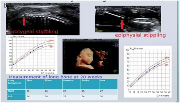
d)X-linked chondrodysplasia punctata: This is a rarer skeletal dysplasia with mild degree of bone shortening.The most striking feature is Binder facies on ultrasound imaging which can be seen in early second trimester.Subsequent prognosis is favorable with mild short stature, no pulmonary hypoplasia or systemic involvement.

If femur length is < -4SD , the most likely diagnosis is a skeletal dysplasia, possibly a lethal one.Other differentials in such a scenario could be conditions with limb reduction defects eg.Focal femoral hypoplasia, Robert’s syndrome, phocomelia, etc.Most of the skeletal dysplasia with such a degree of shortening present in first or early second trimester.Some of the conditions in this category are as follows.

a)Thanatophoric dysplasia:This is a lethal SKD caused by de-novo mutations in FGFR3 gene.It manifests in first or early second trimester with very short limbs, short narrow thorax , a relatively large head and associated hydrops or increased NT.Two distinct phenotypes arerecognised, type I with bent femurs(telephone receiver shaped) and normal shape of head, and type II with clover leaf shaped skull and normal contour of femur.Ventriculomegaly, migrational abnormalities of brain and rarely cardiac, renal defects are also reported.Prognosis is poor, however recurrence risk is low.


  1. b) Osteogenesis imperfecta type II: : A lethal skeletal dysplasia which is characterisedby poor skeletal mineralisation ,multiple fractures and bending of long bones and ribs.It may present in first or second trimester of pregnancy.Unique features are increasedcompressibility of skull, improved visibility of intracranial structures and acute angulation of tubular bones.Majority of cases are due to de-novo mutations in COL1A1 and COL1A2 genes, however many autosomal recessive forms with mutations in various genes are also known.Genetic testing is using Next generation sequencing based exome sequencing or multigene panel testing.

c)Short rib thoracic dysplasias: These are a group of genetically heterogenous skeletal dysplasiasdue to mutations in various genes involved in primary cilia formation and function.all characterised by extremely short ribs and narrow thorax, with or without polydactyly and other visceral malformations.Presentation may be in first trimester or early second trimester.Genetic testing is using Next generation sequencing based exome sequencing or multigene panel.

d) Achondrogenesis: This is a skeletal dysplasia with extremely short long bones, relatively large head and ossification abnormalities of the skull, spine and pelvis.Three types characterised, type 1a,1b and type 2, these being caused by mutations in TRIP11, SLCA26A2 and COL2A1 genes respectively.inheritance is autosomal recessive in type 1a & 1b, and autosomal dominant due to de-novo mutations in type 2.Due to extreme limb shortening, these are usually reconsidered in first trimester ultrasound, and often have associated hydrops.Genetic testing is using Next generation sequencing based exome sequencing or multigene panel.


2.Bent bones: Certain SKDs present with bent bones on ultrasound, most often along with concomitant shortening.Of these, Osteogenesis imperfecta and thanatophoric dysplasia are themost common types.In addition, certain other SKDs also have bent bones as a prominent feature.Campomelic dysplasia is one such SKD that usually has mild bending of bilateral femurs.Degree of shortening is not significant in this condition, usually between -2 to -4SD.The most commonly involved bones are femur, humerus and tibia.In addition, some unique findings can be appreciated, in form of hypoplastic scapula, 11 pair of ribs, micrognathia and sex reversal/genital ambiguity in male fetuses.It is caused by mutations in SOX9 genes, and is an autosomal dominant condition.Perinatal lethality may occur in few cases due to laryngomalacia.However, most patients would survive into postnatal period with short stature and long bone bending of varying degrees.

3.Hydrops or increased NT: Some of the lethal skeletal dysplasias can present with increased NT or grossly hydropic appearance.These include achondrogenesis, atelosteogenesis, short rib thoracic dysplasias, osteogenesis imperfecta and thanatophoric dysplasia.


4.Syndromic disorders with prominent skeletal involvement: In certain genetic syndromes, skeletal involvement with shortening/other abnormalities of bones is a predominant finding.These are also grouped under genetic disorders of theSkeleton as per the Nosology, 2019.These mimic the skeletal dysplasia on antenatal ultrasound, and can be differentiated based on the additional findings and./or genetic testing

a)Robinow syndrome: This is a condition with prominent mesomelic shortening of long bones, more significantly involving the upper limbs.In addition, other characteristic features can be appreciated in form of vertebral segmentation defects and facial dysmorphism.Postnatal outcome is fair, with short stature being the primary morbidity in most cases.

  1. b)Oro-facial digital syndrome: These are a group of conditions characterised by polydactyly, variable oro-facial clefts, tongue lobulation or hamartomas with or without malformations involving brain and kidneys.They overlap with the short rib thoracic dysplasias, and may not be differentiated on antenatal ultrasound without genetic testing.postnatal prognosis is usually guarded due to multisystemic involvement.
  2. c)Oto-palato-digital syndrome(OPD): OPD 1 & 2 are X-linked disorders caused by mutations in the FLNA genes.They are characterised by micrognathia, cleft palate, hand and feet deformities, with or without visceral malformations.Postnatal prognosis is guarded, especially in males
  3. d)Microcephalic osteodysplastic dwarfism: These are a group of disorders usually associated with symmetric bone shortening, overall fetal growth restriction and prominent microcephaly.Postnatal prognosis is variable as per the individual disorder, however usually extreme short stature and microcephaly are present.


Approach to ultrasound for genetic diagnoses

Antenatal ultrasound can be used to arrive at a presumptive genetic diagnoses of a specific SKD by paying attention to various findings as mentioned in table 2.Briefly, besides assessment of lethal/non lethal condition, assessment of degree of shortening, pattern of limb segment involvement ,mineralisation, associated malformations, timing of diagnoses and other findings as discussed in individual conditions above; can help in suspecting a specific SKD.This can further be used to counsel families and perform the specific genetic investigation.Consultation with a Clinical geneticist should be sought for pattern recognition and genetic differential diagnoses, and for decision regarding the best laboratory testing approach ,individualised for the patient.


Role of Postmortem examination

A definitive ultrasound diagnosis is possible in only 35-68% cases.Postmortem examination of the fetus including external dysmorphology, radiographs, internal dissection and tissue histopathology is very important for detailed phenotype delineation.This evaluation should be done in a multidisciplinary manner involving a clinical geneticists and a perinatal pathologist.The post-mortem findings also help in reverse phenotyping and geneotype-phenotype correlation after the genetic test results are available.


Genetic testing for Skeletal dysplasias

SKDs are genetically heterogeneous, and mutations in more than 450 different genes can present with abnormalities of theSkeletonAll these are Mendelian/Monogenic diseases, and cannot be diagnosed by cytogenetic tests like karyotype or chromosomal microarray.Few of the SKDs like Achondroplasia are caused by one specific mutation in majority of patients, and this can be diagnosed using a simple Polymerase chain reaction in a targeted manner.Similarly, campomelic dysplasia and Ellis Van crevald diseases are genetically homogenous and targeted gene sequencing can be done for these using conventional Sanger sequencing.However, for most other SKDs, genetic heterogeneity is the norm, and hence the best diagnostic approach is to use a Next generation sequencing based Exome sequencing or Skeletal dysplasia gene panel.This is especially relevant in the prenatal period, when the specific diagnosis may not be certain.NGS based testing is now available in various laboratory across India at a reasonable cost.The turnaround time ranges from 2-4 weeks, and this may limit its utility in the prenatal period.Also, this testing is likely to reveal variants of uncertain significance and other confusing results like incidental or secondary findings.Hence, this should be performed in collaboration with a Clinical geneticist who should be involved in the Pre- test and post-test counseling of the patients.Besides, confirmation of a specific genetic diagnosis, identification of the causative mutation helps in providing accurate recurrence risks and opportunity for early, definitive prenatal testing or preimplantation genetic testing for these families.


Conclusions

SKDs are a large group of genetic disorders presenting the fetal life with skeletal abnormalities.These disorders have important implications for postnatal outcome both in terms of short stature and various other disabling morbidities, a significant proportion being perinatal lethal.A multidisciplinary approach towards ultrasound diagnosis and laboratory genetic testing with involvement of a clinical geneticist is essential for management of pregnancies affected with a SKD.In view of significant recurrence risk for such families, genetic counselling is an important component of the clinical protocol.

Table 1. List of some common prenatally detected Skeletal dysplasias

Lethal Skeletal Dysplasias Gene involved
Thanatophoric dysplasia FGFR3
Homozygous achondroplasia FGFR3
Achondrogenesis COL2A1,TRIP11,SLC26A2
Short rib thoracic dysplasias WDR60, WDR35,WDR34, TCTEX1D2,SRTD12,NEK1,KIAA0753,KIAA0586,INTU,IFT81,IFT52,IFT43,IFT172,IFT140,DYNC2LI1,CEP120
Campomelic dysplasia SOX9
Hypophosphatasia ALPL
Jeune’s asphyxiating thoracic dystrophy DYNC2H1IFT80, WDR19,TTC21B, SRTD1
Atelosteogenesis- I,II,III FLNB,SLC26A2
Diastrophic dysplasia SLC26A2
Fibrochondrogenesis COL11A1,COL11A2
Non Lethal Skeletal Dysplasias
Achondroplasia FGFR3
Jeune’s asphyxiating thoracic dystrophy DYNC2H1IFT80, WDR19,TTC21B, SRTD1
Ellis Van Crevald syndrome EVC1,EVC2
Osteogenesis imperfecta II COL1A1,COL1A2,CRTAP, P3H1,
SPARC,TENT5A,KDELR2,BMP1,
TMEM38B,IFITM5,CREB3L1,
SERPINH1,WNT1,SP7,PPIB,MESD,
SERPINF1,FKBP10,MBTPS2
COL2A1,TRIP11,SLC26A2
WDR60, WDR35,WDR34, TCTEX1D2,SRTD12,NEK1,
KIAA0753,KIAA0586,INTU,IFT81
IFT52,IFT43,IFT172,IFT140
DYNC2LI1,CEP120
SOX9
ALPL
DYNC2H1IFT80, WDR19,TTC21B
, SRTD1
FLNB,SLC26A2
SLC26A2
COL11A1,COL11A2
Non Lethal Skeletal Dysplasias
FGFR3
DYNC2H1IFT80, WDR19,TTC21B
, SRTD1
EVC1,EVC2
COL1A1,COL1A2,CRTAP, P3H1,
SPARC,TENT5A,KDELR2,BMP1,
TMEM38B,IFITM5,CREB3L1,
SERPINH1,WNT1,SP7,PPIB,MESD,
SERPINF1,FKBP10,MBTPS2
Metatropic dysplasia TRPV4
Spondyloepiphyseal dysplasia congenita COL2A1
Campomelic dysplasia SOX9
Opismodysplasia INPPL1
Diastrophic dysplasia SLC26A2
Kneist Dysplasia COL2A1


Table 2: Distinguishing ultrasound findings in some Skeletal dysplasias

Bent bones
Osteogenesis imperfecta
Thanatophoric dysplasia type 1
Campomelic dysplasia
Kyphomelic dysplasia
StuveWeidemann syndrome
Poor mineralisationion
Osteogenesis imperfecta
Hypophosphatasia
Achondrogenesis
Atelosteogensis
Large head
Thanatophoric dysplasia
Achondrogenesis
Achondroplasia
Cleft lip/palate
Short rib thoracic dysplsias
Oto-palatodigital syndrome
Oro-facial digital syndrome
Atelosteogenesis
Diastrophic dysplasia
Renal/brain/cardiac malformations
Short rib thoracic dysplasias
Oto-palatodigital syndrome
Oro-facial digital syndrome
Hitch Hiker thumb
Atelosteogenesis
Diastrophic dysplasia
Vertebral segmentation defect
Robinow syndrome


Figure:

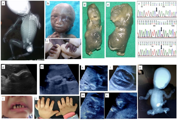
1a-c: Fetus with X linked chondrodysplasia punctata; 1a: Radiograph showing epiphyseal stippling at multiple joints, 1b:Binder facies , 1c: Brachytelephalyngy

1d-f: week Fetus with Achondrogenesis type; 1b,e: Hydrops, extremely short limbs; 1f: Sanger sequences of and parents with c.532C>T, p.Arg178* mutation in exon 2 of SLC26A2 gene

1g-j: Ultrasound and Neonatal photo of fetus with Ellis Van Crevald syndrome; 1g: Depressed nasal bridge and tall forehead on sagittal facial profile, 1h:Postaxial polydactyly , 1i: Dental dysplasia, 1j: Nail dysplasia

1k-o:Fetus with Osteogenesis Imperfecta; 1k: Coronal image of chest showing narrow and bell shaped thorax, 1l: Head showing increasing visibility of intracranial structures, 1m: narrow thorax with short,irregular ribs, 1n: Acutely bent long bone, io: Fetal radiograph showing poor bomineralisationion, short, irregular ribs and short ,irregular long bones with acute angulations

gentic

Postnatal evaluation and management of a case of suspected skeletal dysplasia

Introduction: Skeletal dysplasia are a heterogenous group of disorders which affect the natural development of skeletal system and manifest as abnormalities of limbs, chest, skull or spine. These are genetic diseases which result in abnormal formation of chondro-osseous tissues. Their prevalence varies from 1:5000 to 1:3000 births with such wide variation primarily because of incomplete evaluation of still births and early neonatal deaths in several studies (1). These disorders contribute to a significant proportion of still births and early neonatal deaths, however several of them have better prognosis with survival well into adulthood. They have a varied clinical presentation and can be diagnosed at various ages due to this variable presentation (2). A comprehensive knowledge of these disorders is required for timely suspicion, complete examination, appropriate testing and diagnosis. Early and correct diagnosis is essential as most of these cases are lethal and a diagnosis helps in subsequent pregnancies.

Clinical presentation: Skeletal dysplasias may present in either of the following four presentations: (I) Still birth with antenatal suspicion (growth retardation, short bones, vertebral and facial anomalies) with/without antenatal diagnosis (II) still birth undiagnosed with postnatal findings of shorening, limb, chest or facial anomalies (III) neonate with shortening of trunk and/or limbs, facial anomalies, respiratory insufficiency, fractures (IV) infancy to adulthood with short stature and limb or spine deformities, fractures, eye/ear/dental abnormalities.

Clinical evaluation of a case of suspected skeletal dysplasia: Skeletel dysplasia is a very broad group involving host of disorders. The ones which present during neonatal period are listed in table 1 along with their features. A complete head to toe examination is required to find out all the abnormalities which can help in diagnosis. Skull should be examined for shape (barchycephaly, clover leaf, craniosynostosis), facial abnormal features like hypo/hypertelorism, cleft lip/palate, dentition anomalies, thorax should be examined for size, signs of respiratory insufficiency, spine should examined for kyphoscoliosis, limbs examined for shortening and contractures, site of shortening like proximal (rhizomelia), middle (mesomelia), distal (acromelia). Femur length-foot length ration less than 1 suggests skeletal dysplasia. Table 2 illustrates these clinical features along with their associated causes.

Radiological evaluation of a case of suspected skeletal dysplasia: A comprehensive and detailed skeletal survey is essential in aiding in diagnoses (3,4). Survey must include AP & lateral views of skull and thoracolumbar spine, AP views of chest, pelvis, one upper and lower limb, left hand (for bone age). The axial and appendicular skeleton must be properly examined for abnormalities like workman bones, thick skull, degree of ossification, platyspondyly (flat vertebral body), coronal clefts (radiolucent band running through at least one vertebral body), abnormal vertebral bodies like central beaking in Morquio’s disease and posterior hump shaped in SEDT. In appendicular skeleton, the site of affection (epiphyses, metaphases or diaphyses), type of limb shortening (rhizomelia, mesomelia, acromelia). The aetiologies associated with these findings are elaborated in table 2.

Postnatal management: After the delivery of a case of suspected skeletal dysplasia, initial stabilisation is very crucial which is to be followed by multi-disciplinary approach to diagnosis and ongoing care. Table 3 depicts the summarised care of such a baby. It is very essential to obtain samples for genetic tests for diagnosis as many of these disorders have poor prognosis and antenatal diagnosis in future pregnancies is crucial.

Conclusion: Skeletal dysplasia is a group of disorders with varied presentation and a strong clinical suspicion is necessary for timely diagnosis as outcomes are not very good. A multidisciplinary team approach is necessary to ensure a proper quality of life to survivors. Appropriate timely antenatal diagnosis at this point is primarily the mode of management. Future research on gene therapy and other treatments are required for ensuring better survival and quality of life to these individuals.

Category Disease name Clinical and radiologic features Genes involved, inheritance and prognosis
FGFR3 group Thanatropic dysplasia
  • Narrow thorax with pulmonary hypoplasia
  • Short extremeties, normal trunk length
  • Cloverleaf skull, telephone receiver femurs
  • 4p16
  • AD
  • Lethal
Achondroplasia
  • Short extremeties, rhizomelia, normal trunk lengt
  • Macrocephaly, hydrocephalus
  • Trident hand, Hypotonia
  • Foramen magnum stenosis
  • Recurrent otitis media
  • Normal intelligence
  • 4p16
  • AD
  • Excellent
Hypochondroplasia Similar to achondroplasia, milder severity, later onset in second decade
  • 4p16, AD
  • Excellent
Type II collagen group Achondrogenesis type II
  • Hydrops, short trunk, prominent abdomen
  • Severe limb shortening
  • Severely retarded bone ossification
  • 12q 13 (COL 2A1)
  • AD, Lethal
Hypochondrogenesis
  • Similar to achondrogenesis but milder shortening and better ossification
  • Most die within first 3 months from respiratory insufficiency
  • 12q 13 (COL 2A1)
  • AD, poor
Kniest dysplasia
  • Thoracic kyphoscoliosis, lumbar lordosis
  • Platyspondyly, anterior vertebral wedging, coronal clefts
  • Flat mid face, depressed nasal bridge, cleft palate
  • Joint contractures, delayed ossification and deformation of epiphyses
  • Myopia, retinal detachement
  • Chronic otitis media, hearing loss
  • 12q 13 (COL 2A1)
  • AD
  • Fair to good
Spondyloepiphyseal dysplasia congenita
  • Similar to Kniest dysplasia but milder
  • Normal long bones
  • Coronal clefts absent
  • 12q 13 (COL 2A1), AD
  • Fair to good
Stickler syndrome
  • Midface hypoplasia, small upturned nose, cleft palate, Pierre Robin sequence
  • Joint hyper mobility
  • Vertebral coronal clefts, widened epiphyses
  • Myopia, cataracts, hearing loss
  • 12q 13 (COL 2A1),
  • AD
  • Good
Short rib dysplasia group Ellis van Crevald syndrome
  • Narrow thorax, dental anomalies
  • Polydactyly, rhizomelia, hypoplastic nails
  • Congenital heart disease
  • 4p 16 (EVC1&2), AR
  • Fair to good
Asphyxiating thoracic dysplasia (Jeune)
  • Narrow thorax with respiratory insufficiency
  • Polydactyly, short hands and feet
  • Metaphysical irregularities, short middle & distal phalanges
  • Chronic kidney disease, pancreatic and hepatic fibrosis
  • Hirschsprung disease, multiple gingival frenulae, hydrocephalus
  • 3q 24-26 (IFT80), 15q 13
  • AR
  • Fair
Decreased bone density group Osteogenesis imperfecta type I-IX
  • Type II and III evident at birth, others variable age group presentation frm infancy to adulthood
  • Type II most severe with multiple fractures and high lethality, others with variable fracture rates and short stature severity
  • Blue sclera in types I to III, hearing loss in types I and III
  • I, IV,V: AD
  • II & III: AD or AR
  • VI-IX: AR
  • I to IV: COL1A1 or 2
Defective mineralization group Hypophosphatasia

  • Perinatal lethal
  • Infantile
  • Severe shortening, respiratory failure, seizures
  • Undermineralization, rachicitic metaphases, craniosynostosis
  • Short stature, premature loss of deciduous teeth, symptomatic hypercalcemia
  • 1p36 (TNAP), AR, lethal
  • 1p36 (TNAP), AR, variable

Table 1: Skeletal dysplasia which present during neonatal period. Abbreviations: AD autosomal dominant, AR autosomal recessive.

Part of body Clinical or radiological feature Possible ethology
SKULL Macrocephaly/ Microcephaly TD, achondroplasia, JLS/ CDP
Irregular skull shape TD (cloverleaf), CDP, JLS
Hydrocephalus CD, Oi type 2, osteoporosis
Normal size

  • Partial absent bones
  • Total absent bones
  • Hypoechogenic bones
CCD, OI type 1 & 2CCD

CCD, OI type 2, achondrogenesis

Wormian bones/ Thick skull CCD, PD, OI, hypophosphatasia, Osteopetrosis, CFD
FACE Facial anomalies (micrognathia, hypo/hypertelorism, frontal bossing CDP, achondroplasia, CD, cleidocranial dysplasia, DD, OI type 1, SEDC
Cleft lip or palate/ Gingival frenulae SRPS, CD/EVS
Eye abnormalities:

  • Orbit anomaly/Ctataract
CCD/CDP
Teeth

  • Natal teeth
  • Supernumerary teeth
  • Dentinogenesis imperfecta
  • Hypoplasia of dental cementum
EVSCCDOIHypophosphatasia
THORAX Narrow

  • Short horizontal ribs
  • Short ribs, 1 or more absent
  • Hypoechogenic ribs with fractures
  • Hypoechogenic ribs without fractures
Osteopetrosis, CCD, EVSTD, CCD, JLSOI type 1&3TD, achondrogenesis, OI type 3
Short barrel shaped JLS, SEDC
LIMBS Normal limb echogenecity with severe shortening

  • Rhizomelia with bowing
  • Rhizomelia with straight bones
  • Mesomelia with bowing
  • Mesomelia with straight bones
  • Rhizomesomelia with bowing
  • Rhizomesomelia with straight bones
Amelia, osteoporosis, SRPS, CCD, EVS
TD, CCD, JLS
OI type 1&3
TD, achondrogenesis 1A & 1B, OI type 3
HC, JLS, OI type 3, SRPS, SEDC
DD, FHUFS
Normal limb echogenecity with mild to moderate shortening

  • Rhizomelia
  • Mesomelia
  • Rhizomesomelia
EVSMesomelic dysplasia, SEDCOI type 1, SRPS, SEDC
Decreased limb bone echogeenicity

  • Short limbs
  • Normal length limbs
Achondrogenesis, OI type 1 & 2
OI type 2
Hypoplastic nails EVS, CDP
SPINE Platyspondyly MD, Morquio syndrome, OI, TD, KD
Coronal cleft CDP, MD, KD

Table 2: Clinical features in case of skeletal dysplasia. Abbreviations: OI osteogenesis imperfecta, TD thanatropic dysplasia, JLS Jarcho-Levin syndrome, CDP chondrodysplasia punctata, CD campomelic dysplasia, CCD cleidocranial dysplasia, CDP chondrodysplasia punctata, DD diastrophic dysplasia, SEDC spondyloepiphyseal dysplasia congenita, SRPS short rib polydactyly syndrome, EVS Ellis van Creveld syndrome, HC Hypochondroplasia, FHUFS femoral hypoplasia-unusual face syndrome, MD metatrophic dysplasia, KD Kniest dysplasia, CFD craniofacial fibrous dysplasia, PD pyknodysostosis

Arm of treatment Components
Initial stabilisation Airway and breathing:

  • Respiratory insufficiency requiring invasive or non invasive ventilation
  • Xray to assess pulmonary hypoplasia and thorax dimensions

Circulation:

  • Monitor BP, CFT, echo to assess cardiac disease and PPHN
  • Shock management, management of pulmonary hypertension

Neurological:

Seizures management with AED

  • Apnea: mechanical ventilation

Others:

Handling: if suspicion of OI then gentle handling and supportive care to prevent fractures

  • Skeletal survey: all necessary X-rays as stated above radiological evaluation section
  • Nutrition managenet: pareneteral nutrition if sick and unable to take enterally
  • Fluid and electrolyte balance: as these babies are IUGR, may have associated PPHN, shock, renal disease
Multidisciplinary approach to diagnosis
  • Intensivist: to manage the components of survival as stated above in initial stabilisation
  • Radiologist: interpretation of skeletal survey and aiding in diagnosis
  • Medical geneticist: to aid in understanding the mode of inheritance (family tree), required genetic tests, aiding in diagnosis and counselling
  • Cardiologist: to aid in diagnosis of any cardiac disease and management of shock and/or PPHN
  • Pulmonologist: management of respiratory insufficiency and management of chronic lung disease
  • Nephrologist: management of CKD in select cases
  • Neurologist: management of seizures, neurodevelopment assessment
  • Orthopedics: management of fractures, correction of deformities
  • Physiotherapist and Occupation therapist
  • Ophthalmologist and ENT: eye and ear problems as discussed
  • Nutritionist: to ensure adequate growth
  • Psychologist: to provide support to family
  • Dentist: to manage dentition associated problems
  • Neurogsurgeon: problems like cord compression, hydrocephalus
Specific therapies
  • OI – bisphosphonates, growth hormone, teriparatide
  • Hypophosphatasia – asfotase alfa
Postmortem analysis Include the following:

  • Photographs
  • Skeletal survey
  • Skin and tissue biopsies

Table 3: Postnatal management in case of suspected skeletal dysplasia. Abbreviations: BP blood pressure, CFT capillary refill time, PPHN persistent pulmonary hypertension, OI osteogenesis imperfecta, CKD chronic kidney disease.

References:

  1. Andreas Zankl.Genetics of Bone Biology and Skeletal Disease (Second Edition), 2018
  2. Seth J Langston, Deborah Krakow, Alison Chu. Revisiting Skeletal Dysplasias in the Newborn. Neoreviews 2021 Apr;22(4):e216-e229.
  3. G R Mortier. The diagnosis of skeletal dysplasias: a multidisciplinary approach. Eur J Radiol 2001 Dec;40(3):161-7.
  4. D Krakow. Skeletal Dysplasias. Clin Perinatol. 2015 June ; 42(2): 301–319.