Fetal growth and monitoring has always been a subject of tremendous interest
for the obstetricians ,Fetal medicine specialists, neonatologists and not to forget
-the parents!
[envira-gallery id=”307822″]
Research from various parts of the world has provided the much needed insight
in this aspect however published work from Indian quarters has always been a
sore point.This is especially when we have some researchers arguing that
growth is ethnicity specific with an equally vociferous disagreement.
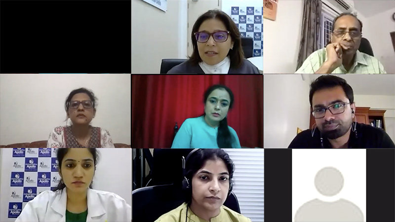
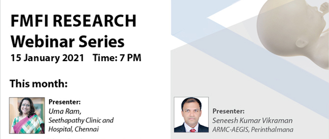
We were however fortunate to have Dr Uma Ram and Dr Seneesh KV for the
FMFI Research Webinar on 15.01.21 who have initiated work in this field.
Dr Uma Ram presented her work on how the Diabetic foetuses have increased
subcutaneous fat at the midtrimester scan, predating the clinical diagnosis of
GDM. She also shared that these studies are now being validated as prospective
studies.
An interesting discussion by the chairpersons- Dr S Suresh, Dr Ashok
Khurana and Dr Anita Kaul was whether these findings could be integrated in
an algorithm to initiate early maternal nutrition therapy.
Dr Seneesh showcased his study, prospectively comparing different available
antenatal and postnatal growth charts.
The concluding remarks by the chaipersons emphasised the need of following up every fetus by the
drop/increase in their centiles irrespective of the growth charts that are used. In
addition, multivessel Dopplers added significant value in the decision of
delivery of a fetus that shows declining centiles.
It was a stimulating session and pushed us to think beyond the routine where a
simple additional measurement from a standard fetal plane could potentially
change the course of maternal complications. The hard work put in a
prospective study whilst doing routine clinical work to answer questions that we
come across daily but brush aside due to lack of time was equally invigorating.
We hope to collate many more such research work by Indian authors in the
times to come.